Ventricular Septal Defect Closure (VSD) - Signs, Symptoms, Treatment

Ventricular Septal Defect Closure (VSD) Details

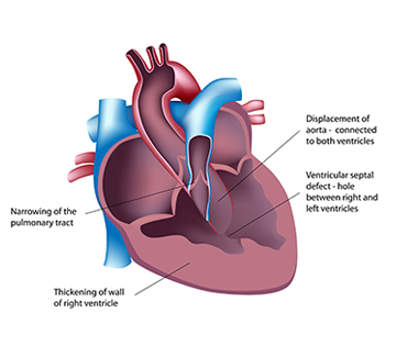
Your heart is a wonderful organ. It beats constantly 24/7 to ensure the supply of oxygen rich blood to all the parts of your body. It performs this activity tirelessly throughout your life. The heart is divided into four chambers; the upper chambers are called atrium and lower chambers are called ventricles. The wall dividing the left and right side of the heart is called septa. At times there is a small opening in the septa which does not keep the ventricles completely separated from each other. This is a serious condition and requires medical attention. This can also result in mixing of oxygenated and deoxygenated blood, thus reducing the oxygen carrying capacity of the blood. This condition requires a minimally invasive technique to close the opening.

Get a Free Personalized quote for Ventricular Septal Defect Closure (VSD)

(jpeg, jpg, doc, docx, pdf)

Before Ventricular Septal Defect Closure (VSD)

You will need to do all types of preliminary tests to confirm the exact location of the opening, its size etc. Your doctor will also carefully run through your reports of ECG and angiography. These will help him understand any abnormality in your heart beat or if there are chances for you to develop any other major cardiac problem in the near future. You might be put under special medication to prepare your body for the procedure and will be asked to stop drugs which can hinder the surgery. It is always good to share your complete medical history with the doctor and arrive for the surgery on an empty stomach.

How it is performed

You will be under anesthesia and will not feel any pain or discomfort. Your doctor will make an incision in your chest cavity or groin area, to try to get into a blood vessel that can lead to your heart. All this is monitored with the help of graphs or images which are being taken regularly and are being viewed on a screen by your doctor. After the vessel has been identified your doctor will introduce a guide tube in the blood vessel and will make it reach the heart. To close the defect, your doctor will enter the left ventricle and will make the guide tube travel through the ventricular opening to the right ventricle. After it has reached the right ventricle, your doctor will put a catheter in the guide tube and will make it reach the right ventricle. After reaching the right ventricle, the first portion of a medical grade mesh is expanded in the form of a disc and is then adjusted on the opening. It is pulled slightly backward (towards left ventricle) to make sure of a good fit. Now it is time to expand the mesh in the left ventricle. After it has expanded at both the ends, it is fixed and the catheter and guide tube are withdrawn. The mesh behaves like the ventricular wall and in some time gets covered by cardiac tissue.

Recovery

This is a minimally invasive procedure and does not require long hospitalization. You can go home after your surgeon feels satisfied with your recovery. You need to make small changes to your lifestyle initially to set everything as expected. Normal life can be resumed as early as possible. It is advised to avoid tasks that demand your heart to strain.

Potential Risks

The procedure is percutaneous and does not have any serious threat associated with it. It is also a gold standard for the surgery. The only problem that has been associated in some individuals is relocation of device or the device has become loose etc. All these risks can be managed if your surgeon insures the most accurate fit of the device and by holding it in place securely and tightly. You can avoid majority of complications by following the instructions given by your doctor.a

Cost for Ventricular Septal Defect Closure (VSD)

India from $3,500
Turkey from $15,000
Egypt from $13,000

Ventricular Septal Defect Closure (VSD) Hospitals

Free Enquiry