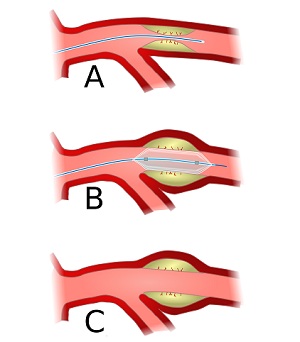
Coronary angioplasty, also termed percutaneous coronary intervention, is an elaborate process that involves making a small incision in the groin or arm to get access to the artery. In the process, the cardiologist guides a thin wire through the small incision made to reach the blocked artery. Once the wire comes in contact with the blocked artery, a deflated balloon is then inflated that eventually helps widen the artery, leading to an improved blood flow.
During the angioplasty procedure, plaque removers are used to eliminate deposited plaque that accumulates on the walls of the arteries. Contemporary devices such as stents are used to open the blockage in the artery. In this case, a stent is permanently fixed. Once, four to five hours have passed post the coronary angioplasty procedure, the catheter and the wire are removed. Read more.
The entire coronary angioplasty procedure takes a maximum of two hours. At the time of the procedure, medication is administered to patients to keep them awake, but they feel drowsy. The major purpose of coronary angioplasty is to open the narrowed blood vessels. This helps enhance the flow of blood to the heart. The procedure helps reduce the risk of heart attack to minimise the symptoms of angina. This procedure also helps figure out the progress of coronary artery disease.