How Much Does Brain Tumor Surgery Cost in India?

  • Brain Tumor surgery cost in India starts from USD 4800 (craniotomy), which is less than one-third of the price for the same procedure in the USA, European countries and other nations. 
  • The duration of stay in the hospital is 4-6 days and in India, approximately 3 weeks.
  • The treatment plan is concluded after complete evaluation of the patients for determining the size, location and type of tumor. 
  • With new and improved surgical technologies, brain tumor surgery has become more safe and effective in treating various kinds of brain abnormalities.
Brain Tumor surgery cost

Brain Tumor Surgery Cost in India

India is one of the most sought after destinations for medical related travel, mainly due to the remarkably low prices for high quality treatment at the top hospitals.

The cost of brain tumor removal surgery, performed by experienced and renowned neurosurgeon, typically ranges from USD 4800 to USD 9000. This cost is subject to variation based on multiple factors.

Some of these parameters that may influence the overall cost of brain tumor treatment include:

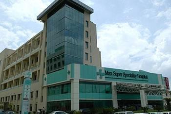
  • The choice of hospital: Location and standard of quality offered by the hospital impacts the cost of healthcare package they offer. Internationally accredited brain tumor hospitals in India, such as Fortis Research Memorial Institute (FMRI), Indraprastha Apollo, Max hospital, Kokilaben Dhirubhai Ambani Hospital and others offer world-class medical services to the patients and their attendants. These are located in major cities, which are easily accessible through international airports (connected to all major airlines of the world) and other modes commute.
  • Surgeon’s experience: India is home to several renowned surgeons. The qualifications and years of experience in their field of expertise also varies the surgeon’s fee in the cost package. The best brain tumor surgeons in India are trained in advanced techniques from the top medical institutions and have a vast experience of treating various kinds of brain abnormalities. Read in detail about these doctors in the article 15 Best Neurosurgeons in India.
  • Type of surgery and technology used: The hospitals in Delhi (NCR), Mumbai, Bangalore, Chennai and other cities are equipped with high-end technology and modular Operation Theatres to provide the best treatment to brain cancer patients. Minimally invasive technologies such as Robotic arm system increases the precision and minimizes the human errors during the surgery. The price may increase if the patient opts for such technology.
  • Certain kinds of brain tumor can be treated with radiosurgery techniques, such as GammaKnife and CyberKnife. It is important to note that these are radiation therapy and not surgical procedures. Radiosurgery is an advanced cancer treatment technique which allows patients to undergo a radiation therapy with high precision and accuracy. It enables the precise targeting of cancer cells, with minimal or no damage to surrounding healthy tissues. Sometimes, GammaKnife and CyberKnife are preferred over conventional surgery methods. This is will be determined by the doctor based on type, location and size of tumour, and patient’s overall health condition.

For more details, read CyberKnife treatment in India and GammaKnife treatment in India.

  • Length of hospital stay: Normally, the patient will have to be hospitalized for 4-6 days, but this may vary depending on patient’s medical condition, speed of recovery and risk of complications. The room charges will expensed on number of days of stay required.
  • Room category: The hospitals offer a variety of rooms for post-surgery hospitalization, including single, twin sharing, triple sharing and deluxe. The cost is different for each category room.
  • Other miscellaneous: Addition tests or procedures, if needed, also add up to the total cost of brain cancer treatment in India.

Top brain tumor surgery hospitals in India offer cost effective health packages, which enables international patients to save as high as 60-80% money in compared to what they will have to spend in Western countries. To know more details about the cost breakdown and inclusion/exclusion of the package, share your query with us.

Our team of experts will contact at the earliest and address all your questions. We will further connect you to the best neurosurgeons for initial consultation.

Connect with Experts & Get a Free Personalized Quote for Brain Tumor Treatment in India

(jpeg, jpg, doc, docx, pdf)

What is brain tumor?

Brain tumor is a mass of abnormal cells in the brain, that develops due to uncontrolled growth of cells. It may originate in the brain cells only, known as primary brain tumor, or form due to metastasis of cancer cells from another part of body (where the cancer originally started), known as secondary brain tumor. The primary tumor can be benign or malignant (cancerous), and located in any part of the brain. 

Although, the definitive causes of brain cancer is yet unknown, several genetic and environmental factors are attributed to it. 

Types of brain tumor:

There are many types of primary brain tumors that can be classified based on the type of cells they originate from, location of tumor and other factors. They are mainly calssified as glioma, and non-glioma tumor types in adults.

Glioma begin in the glial or supportive tissue of the brain and are the most common type of primary brain tumor. Some common types of gliomas are:

Astrocytomas: The tumors begins in astrocytes, small, star-shaped cells and can grow in any part of the brain or spinal cord. In adults, astrocytomas most commonly arise in the cerebrum, whereas in children, they often occur in the brain stem, the cerebrum and the cerebellum. 

Brain stem gliomas: This type of glioma occurs in the lowest part of the brain, near the stem. The brain stem is responsible for many vital functions of the body. Most brain stem gliomas come under high-grade astrocytomas.

Ependymomas: These tumors often develop in the lining of the ventricles in the brain or in the spinal cord. They are mostly found in childhood and adolescence, but can develop at any age.

Oligodendroglioma: This kind of tumor begins in the cells that produce myelin, the protective covering of the nerves. It usually occurs in the cerebrum and are rare. They grow slowly and normally do not spread to surrounding healthy brain tissue. They are mostly found in middle-aged adults but can occur in people of all ages.

Other types of brain tumors that do not begin in glial tissue are called Non-gliomas. Some of the most common types are:

Medulloblastomas: This kind of brain tumor develop from primitive or developing nerve cells which generally are not found in the body after birth. Medulloblastomas are therefore also referred to as primitive neuroectodermal tumors (PNET). Mostly, they begin in the cerebellum, but may occur in other parts as well. These tumors are most often found in children, more commonly in boys than girls.

Meningioma: These tumors grow from the meninges, or membranes that enclose the brain and spinal cord. They are usually benign. Because these tumors grow very slowly, the brain may be able to adjust to their presence. Meningiomas often grow quite large before they cause symptoms. They occur most often in women between 30 and 50 years of age.

Pineal region tumor: These non-gliomas are typically found in or around the pineal gland, a small organ located near the center of the brain. This tumor can be slow growing (called pineocytoma), or fast growing (called pineoblastoma). As pineal region is in the centre of the brain, it is very difficult to reach, and tumors in the part often cannot be removed surgically.

Craniopharyngioma: This type of tumor occurs in the pituitary gland, located near the hypothalamus. These are usually benign tumors but can be sometimes considered malignant as they may press upon or damage the hypothalamus, which affect certain vital functions of the brain part. They are most often found in children and adolescents.

Schwannoma: It is a rare tumor that develops in the Schwann cells, which produce the myelin that protects the nerve of hearing (acoustic nerve). These tumors are usually benign and mostly occur in adults, twice in women than men. It affects the vestibular nerve in the inner ear, which helps control balance. 

Germ cell tumors: These types of tumors occur in the developing sex cells or germ cells. Germinoma is the most common type of germ cell tumor in the brain.

How can Lyfboat assist you getting Brain Tumor surgery?

World's Most Trusted Medical Advisory & Discount Platform

Lyfboat is a free advisory platform; we do not charge any fees from patients. In fact, we negotiate the price that Indian hospitals offer. In some cases we are able to reduce the cost by negotiating upto 20% of what Hospitals generally offer. We advise the best treatment from the top hospital/surgeon at best price.

What complications can brain tumor cause?

Some people with brain tumor may not show symptoms till the advanced stages, or the symptoms may appear similar to that for other conditions. The signs and symptoms of brain tumor depend on the size, type, and location of the tumor.

Some general symptoms are:

  • Headaches: severe, persistent and worsen while performing activities or in the early morning
  • Seizures
  • Changes in the personality or memory loss
  • Nausea or vomiting
  • Vision problems, such as blurred vision or double vision

Some symptoms that may be specific to the tumor location include:

  • Feeling of pressure or headache near the brain tumor
  • Altered perception of touch or pressure, arm or leg weakness on one side of the body, or confusion with left and right sides of the body – when parietal lobe of the cerebrum is affected
  • Loss of balance and problems with fine motor skills – when cerebellum is affected
  • Changes in cognitive skills and ability to make decisions, including sluggishness, loss of initiative, and weakness in muscle or paralysis – when frontal lobe of the cerebrum is affected
  • Partial or complete loss of vision – when occipital lobe of the cerebrum is affected
  • Lactation (without pregnancy), changes in menstrual cycle, and hair growth on hands and feet in adults – when pituitary tumor is affected
  • Changes in cognitive skills, speech, memory, hearing, or emotional balance, such as sudden aggressiveness and problems comprehending words – when temporal lobe of cerebrum is affected
  • Inability to look in upward direction – when pineal tumor is affected

What is brain tumor surgery?

Brain tumor surgery is one of the main treatment option for removal of abnormal cells from the brain, along with some surrounding tissue. Surgery is usually the first standard treatment recommended for a brain tumor patient and is often the only treatment needed to eliminate a low-grade brain tumor.

A neurosurgeon is the specialist who performs the surgery on the brain and spinal cord. For a brain surgery, the surgeon will have to remove a part of the skull, a procedure called a craniotomy surgery, to access the brain. After the removal of the tumor, the patient’s own bone is used to cover the part that was removed from the skull. 

The surgery can be performed for following purposes:

  • Remove the entire tumor with an aim to treat it
  • Remove a part of the tumor (partial tumor removal surgery) to slow cancer growth and relieve some symptoms
  • To drain excess cerebrospinal fluid accumulated in the brain and relieve the pressure build-up (hydrocephalus)
  • To provide access for administering chemotherapy
  • To perform biopsy for definitive diagnose and staging of brain tumor

The advances in surgical technology for brain tumor removal has improved the outcomes of the treatment. Some of these advances are cortical mapping, enhanced imaging, and fluorescent dyes.

Cortical Brain mapping: This technology allows doctors to locate and identify the regions of the brain that control functions like senses, language, and motor skills. Other techniques that can be used for brain mapping are Direct cortical stimulation, Evoked potentials, Functional MRI, and Intraoperative ultrasound imaging. 

Enhanced imaging devices: These tools are to help surgeon plan and perform the operation. Computer-based techniques, such as Image Guided Surgery (IGS), enables the surgeon to map the location of the tumor with high accuracy. 

Fluorescent dyes: These agents are given by mouth to the patient in the morning before surgery. They are taken up by tumor cells which can then be seen using a special microscope and light during the surgery, that allows visualization of the cells that take up the dye. This helps surgeon is removing the tumor as safely possible.

The best Neuro hospitals in India are equipped with a variety of sophisticated brain surgery techniques and tools. The neurosurgery departments at many of these hospitals are centre of excellence outfitted with high-end technology for brain surgery and patient care. 

Who may need brain tumor surgery?

Surgery is typically the treatment for low-grade tumor especially when it is possible to remove the entire tumor. Some types of brain tumors may grow rapidly, while others have slow growth. This along with several other factors are considered when surgery is recommended.

For patients with higher-grade tumors, surgery is usually the first line of treatment, which is then followed by chemotherapy and radiation therapy.

A multidisciplinary team of doctors, including neurosurgeons, surgical oncologist, radiation oncologist, medical oncologists and other specialists, are involved in designing a treatment plan for a patient.

Brain surgery may not be a suitable option for all patients. It might be possible to use the latest technologies such as GammaKnife and CyberKnife for treating certain inoperable tumors or for patient who can’t undergo surgery due to health risks. 

The brain tumor surgery is recommended as a treatment option depending on the following criteria:

  • The size, grade and type of brain tumor
  • Whether the tumor is putting pressure on the dynamic parts of the brain
  • Whether the tumor has spread to other parts of the body or Central Nervous System
  • Risks and side effects of the surgery
  • Overall health status and personal preferences of the patient

Brain Tumor Surgery

There are different types of brain tumor surgery that can be performed. The type of surgery needed by the patient depends on the size, location and type of tumor. 

The main types of surgery for brain tumor include:

Craniotomy

A craniotomy is the convention surgery for a brain tumour. It can be carried out under a general anaesthetic or as awake craniotomy. With general anaesthesia, the patient remains asleep and won’t feel anything during the surgery.

  • During craniotomy procedure, the neurosurgeon removes a portion of bone from the skull to provide an opening to access the brain for operation.
  • The doctors will use imaging scans before and during the surgery to locate and identify the tumor. These scans are updated into a computer to enable the doctor to access the precise position of the tumour. 
  • The surgeon will use a variety of tools to remove the tumour, including a scalpel or special scissors. 
  • An ultrasound machine may also be to break up the tumour and then the cells and tissues are suctioned out using a device to gently remove the tumour, this is known as ultrasonic aspiration. 
  • A microsurgery involves the use of a special microscope to visualize the brain tissue (stained with dye) to differentiate between tumour and healthy tissue of the brain. A blue fluorescent light may also be used by the surgeon to see the edges of the tumour more clearly. 
  • After the tumour is removed, the surgeon puts the skull bone, called as a flap, back to close the opening in the skull. The bone is secured using small metal brackets and the skin over it is stitched. In most cases, the scar can be easily hid under hair on the scalp.

Awake craniotomy

An awake craniotomy is mostly recommended by the surgeon when the tumour is close to a part of the brain that controls critical functions such as the feeling, speech, or movement. 

Awake craniotomy can be performed in different ways. Some people are conscious and awake for only part of the operation, while others may remain awake the whole surgery. 

For the first kind, the patient is awake for the later part of the operation and will have a general anaesthetic at the beginning. The anaesthetic is gradually reduced so that the patient slowly wakes up for the other part. Then, another general anaesthetic is given for the last part of the operation.

During an awake craniotomy, the patient is asked to do certain tasks so that the medical team check the function of different parts of the brain. For instance, the patient might be asked:

  • To speak – answer the questions 
  • To move a part of body
  • Describe what you can feel

After the tumour is removed, the skull is repaired with the flap and the skin is stitched up.

A local anaesthetic is sued during the operation to numb the skin on the head and other areas that feel pain such as the muscle. There are no pain receptors in the brain itself and therefore it doesn’t feel pain. 

The surgical team may monitor the brain electrical activity during the operation. This is known as neurophysiological monitoring. The technique allows the surgery to be performed safely and more tumour can be removed.

Neuroendoscopy

Neuroendoscopy is a keyhole procedure for brain surgery. The surgeon may remove all or part of a tumour from the fluid filled ventricles of the brain. Neuroendoscopy can be performed to remove the fluid build up in your brain, condition called hydrocephalus. An endoscope is a long tube with a camera attached to one end. The device might be flexible or rigid, depending on the surgical requirements.

During the procedure: 

  • The surgeon will make a Burr hole, a small hole into the skull. 
  • An endoscope is then inserted through this hole. 
  • The surgeon can see what is ahead of the tip of the endoscope through the attached camera to an eyepiece or on a monitor. 
  • Special surgical instruments, such as small forceps and scissors are at the end of the endoscope. The surgeon uses these tools to remove the tumour.

Other surgical procedures include:

VP Shunt surgery: 

This procedure is also used to drain a build up of fluid on your brain, called hydrocephalus. The build up of fluid can be drained during the brain tumor surgery, or as a separate operation.

A shunt is medical device that drain the excess fluid from the ventricles and send it to abdominal cavity or heart chamber, where the fluid will get disposed off naturally. Read about VP Shunt surgery here.

Surgery to administer chemotherapy

Brain surgery can be used to administer chemotherapy into the area of the tumor. This can be done using an implant or through a ventricular access device that is put in the brain. The ventricular access devices allow patient to receive chemotherapy drugs straight into the fluid filled ventricles of the brain. 

Removing a pituitary tumor via the nose

Pituitary is a small gland organ located above the back of the nose. The surgeon may access and remove the pituitary gland tumour via the nose. This process is called as transsphenoidal surgery.

During the procedure, surgeon creates a small cut on the nasal septum, the thin wall of bone and cartilage separating the nostrils. The surgeon inserts special tools through this hole in the septum, to reach up to the pituitary gland and remove the tumor.

It can be performed using an endoscope. The endoscope allows the surgeon to visualize the insides of the nose and pituitary which operating with surgical instruments to remove the tumor.

The surgical techniques used in the brain tumor treatment:

Depending on the requirement, availability of equipment and surgeon’s skills, different types of surgical instruments and techniques can be used to perform brain tumor surgery.

Stereotactic surgery: Stereotaxy is the use of computers and software systems to create a three-dimensional image. This technique provides precise and detailed information about the location, size and structure of a tumor along with its position relative to the other parts of the brain.

The neurosurgeons use stereotaxy to map out the parts of brain and tumor bedore the surgery. This enables the neurosurgeons to plan the operation as well as rehearse or practice prior the procedure. The technique is also used by radiation specialist to plan the delivery of radiation therapy.

Stereotactic surgical techniques can be used for biopsies, resect tumors, implant radiation pellets or other treatments locally. It can be performed with or without a frame. The frameless stereotaxy technique provides a navigational system to the surgeon during the procedure. 

The benefits of Stereotaxy techniques include 

  • Reaching the tumors that are located deep within the brain, such as in the brain stem or thalamus. 
  • Help limit the extent of surgery. The stereotactic systems can project real-time images of the brain structure while the surgery is being performed. 
  • The three-dimensional images allow the surgeon to precisely use scalpel for cutting, the laser beam for vaporization, insert the needle for biopsy, or the suction device for aspiration.

Embolization: This method is aimed at blocking the blood supply to a tumor by cutting off the flow of blood in selected arteries. It is performed prior to surgery. Whether an embolization procedure is necessary or not is determined on the basis of results from an arteriography, an X-ray taken after injecting radiolabeled dye into the circulatory system.

This test helps doctor determine if and which blood vessel may need to be blocked. Surgery is performed soon after it to avoid re-growth of blood vessels. Embolization technique might be used for vascular brain tumors such as meningiomas, meningeal hemangiopericytomas, and glomus jugulare tumors.

Endoscopy: This involves use of a long, narrow, flexible tube with light and camera at one end, called Endoscope, which is inserted through the surgical incision. This instrument provides light and visual access to the surgeon during the operation.

The advantage of this instrument is that it requires relatively small openings, sometimes called keyhole approaches. The neuroendoscope is particularly used for surgery to correct a malfunctioning shunt, remove scar tissue that is blocking a shunt, or remove intra-ventricular tumors. Endoscope can also be used for removal of brain cysts.

Laser surgery: The laser beams can be used during brain tumor surgery to destroy the tumor cells with heat. It may be used in along with, or in place of, a scalpel. Laser instruments can generate immense heat and focused at close range. The heat energy destroys the tumor cells by vaporizing them.

Stereotactic techniques with advanced software systems can be used to direct the laser. Lasers are mainly used in the treatment of deep-seated tumors within the brain, have invaded the skull base, for hard tumors that cannot be removed using suction method, or with tumors that may break apart easily.

Photodynamic laser surgery: This therapy combines the use of laser surgery and a drug that increases the tissue’s sensitivity to light. Before the surgery, the doctors will inject photosensitizing drug into a vein or artery. The dye travels to the tumor through the circulatory system and accumulate within the tumor cells.

During the operation, the tumor cells will appear fluorescent due to the dye and the surgeon can aim a laser precisely at the tumor cells to activate the drug. This activated form the drug is lethal to the tumor cells. However, it is considered a local form of therapy as some parts of the tumor may not be exposed to the light.

Also, there is a risk of swelling in the brain if tumor is near the brain stem and therefore, such brain tumors cannot be treated with this technique.

Ultrasonic aspiration: This method involves the use of ultrasonic sound waves to break or fragment the tumor into small pieces, which can then be aspirated/suctioned out using a device.

The advantage of this technique is that it causes fewer disturbances to the nearby cells or tissue in comparison to other suction devices. It also causes less heating and destruction of normal tissue. This technique is particularly useful for treatment of tumors that are difficult to remove using cautery and suction due to their firmness and location.

Happy Lyfboat Patients

Facebook
Twitter
WhatsApp
Skype
Send via Email

Six Simple Steps

Free Access to the Network

Our medical advisory platform automatically sends your medical query to our network of expert Doctors, working only at top internationally accredited Hospitals.

Direct Communication

You and the medical providers communicate directly via email or your online patient account; our case managers are always available to help you with this.

Get Verified Quotations

You receive medical opinions and cost estimates within 24 to 48 hours via email and your online patient account.

Compare Quotes & Advices

Lyfboat empowers you to compare the medical opinions, and discuss the options with our very own doctors who help answer your questions, and guide you in making informed decisions on the best treatment option.

Get Discounted Costs

We negotiate your final costs, explore available discount offers, and we handle all your logistics including travel, accommodation, transport, and medical co-ordination.

Return Home Fit

Get cured, pay hospital after treatment and return home safe. Our care team is available throughout your journey to good health!

Vanshika Rawat

Written By Vanshika Rawat

Vanshika Rawat is an experienced content developer. She is very knowledgeable in the field of science and healthcare and has worked under brilliant scientists during her higher education. Vanshika obtained her degrees in Masters in Science and Bachelors in Science (Microbiology with Hons.) from renowned institutions - Panjab University and University of Delhi.
Dr. Surbhi Suden

Verified By Dr. Surbhi Suden

Dr. Surbhi Suden is one of the founders of Lyfboat and a doctor with a renowned name in the Medical tourism industry. She has been working with international patients since 2008 and is a deeply committed professional with a long term vision of transforming the current healthcare scenarios.
Free Enquiry